Third Molar Maturation Stages in Correlation with Cervical Vertebra Maturation Stages in a Saudi Sample- Juniper Publishers
Juniper Publishers- Open Access Journal of Dentistry & Oral Health
Abstract
Introduction: Determination of the age is not only of forensic importance, however in orthodontic treatment planning, the relationship between skeletal and dental development are examined to detect the degree of maturity of the patient. To the best of our knowledge; no such study is investigated the relationship between skeletal maturity and the dental development in Saudi population.
Objective and aim: The aim of the present study is to compare the cervical vertebra maturation (CVM) stages method and dental maturity using tooth calcification stages.
Methods: The current study comprised of 405 subjects selected from Orthodontic patients of Saudi origin coming to clinics of the specialized dental centers in western region of Saudi Arabia. Dental age was assessed according to the developmental stages of upper and lower third molars and skeletal maturation according to the cervical vertebrae maturation stage method. Statistical analysis was done using Kruskal-Wallis H, Mann-Whitney U test, Chi-Square test; t-test and Spearman correlation coefficient for inter group comparison.
Result: The females were younger than males in all cervical stages. The CS1-CS2 show the period before the peak of growth, during CS3-CS5 it’s the pubertal growth spurt and CS6 is the period after the peak of the growth. The mean age and standard deviation for cervical stages of CS2, CS3 and CS4 were 12.09±1.72 years, 13.19±1.62 and 14.88±1.52 respectively. The Spearman correlation coefficients between cervical vertebrae and dental maturation were between 0.166 and 0.612, 0.243 and 0.832 for both sexes for upper and lower third molars. The significance levels for all coefficients were the same at 0.01 and 0.05.
Conclusion: The results of this study show that the skeletal maturity increased with the increase in dental ages for both gender. An early rate of skeletal maturation stage was observed in females. This study needs further analysis using a larger sample covering the entire dentition.
Keywords: Cervical vertebra; Maturation; Third molar; Dental age
Introduction
The orthodontic treatment is most favorable and effective during growth and hence the assessment of growth is significant in treatment planning for dental and maxillofacial malocclusion [1,2]. Several features such as body height, weight and sexual characteristics, dental and skeletal development are used to evaluate the pubertal growth. The stage of tooth maturation can determine the dental maturity and many studies have found an association between dental maturity and skeletal maturity At present, analysis of cervical vertebrae is extensively used to assess skeletal maturity due to its reproducibility from a routine diagnostic lateral cephalogram for orthodontic treatment [3,4]. Various authors have noted that the developmental stages of certain teeth show a high correlation with skeletal maturity [5].Yet very few studies [6,7] are done to evaluate the relationship between tooth calcification stage and cervical vertebra maturation stage. The purpose of this present study is to evaluate the correlation between the CVM stages method and dental maturity using tooth calcification stages of third molars.
Methods
The present study consisted of a total of405 patients attending the Orthodontic clinics of the specialized dental centers in western region of Saudi Arabia. All Participants are of only Saudi origin. The Orthopantomogram (OPG) and lateral cephalogram with orthodontic records of 150 boys and 255 girls with age range 9-20 years were selected. Inclusion criteria were:
a. A high-quality digital panoramic radiograph and lateral cephalogram.
b. No history of any dental or medical disease affecting the normal development of third molar teeth.
c. The following Exclusion criteria were considered and those patients are excluded from the investigation.
d. Any congenital tooth anomalies or congenital anomalies of the 2nd, 3rd and 4th cervical vertebrae such as fusion between cervical vertebrae or presence of secondary possible were eliminated.
e. Patients having any systemic diseases that could affect growth (such as nutritional disturbance, endocrine disorders, syndromes, and long-term consumption of medication) were.
Dental maturation
The assessment of dental maturation was done according to the upper and lower third molar teeth calcification stages. The development of teeth was categorized into different groups, ranging from A (least development) to H (complete development) [8].
Skeletal maturation
The Skeletal maturity was evaluated by skeletal age using cervical vertebra maturation (CVM) stage method, assessing the morphology (shape and inferior border concavity) of three cervical vertebrae (C2, C3, and C4) consisting of six maturity stages (C1-C6) presented by Bacetti et al. [9,10].
Assessment of the sample
All OPGs and lateral cephalogram were viewed on the same computer screen. The assessment of stages of cervical vertebra development and tooth formation for each sample was done by one orthodontist without knowing of the age or gender.
Statistical method
Analysis was performed using the Statistical Package SPSS statistic V22.0 (IBM Corporation, New York, USA). Difference in proportion was tested using Kruskal-Wallis H followed by MannWhitney U test for inter group comparison, and Chi-Square tests. Difference in mean was tested using t-test. Molar stages were correlated with cervical vertebra developmental stages using Spearman's correlation coefficient. All statistical tests were twosided, and the significance level was set at p < 0.05.
Result
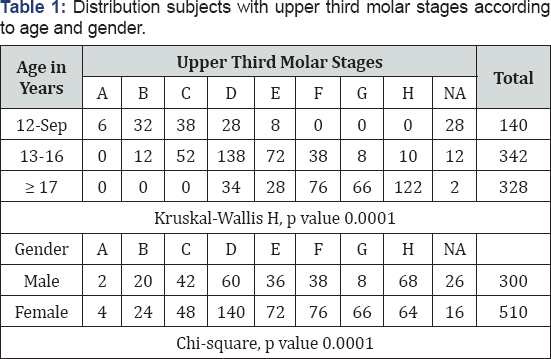
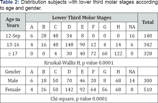
Table 1,2 show the distribution of upper and lower third molar stages according to age and gender. In 9-12 years group the common upper third molar stages were C, B and D respectively, while the common lower third molar stages were C and D then B, there were no F, G and H stages in this age group. In 13-16 years group all third molar stages were present, the most common stage was stage D. In the age group more than 17 years, H stage of third molar was the most common in upper and lower third molars. In the female group of this Saudi sample the most common stage of third molar was stage D, while in the male group the common stages were stage D and H.


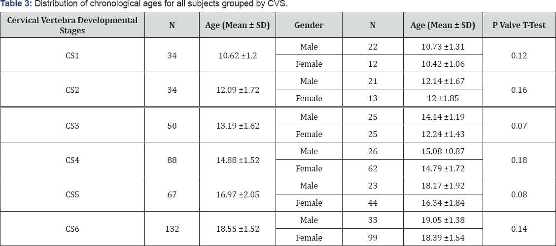
Table 3 shows the distribution of chronological ages for all subjects grouped by cervical vertebra developmental stages. The mean age and standard deviation for cervical stage 3 and 4 were 13.19±1.62 and 14.88±1.52 respectively; the females were younger than males in these cervical stages.

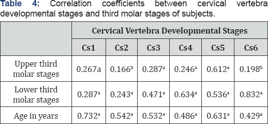
Table 4 shows the correlation coefficients between cervical vertebra developmental stages and third molar stages of subjects in both upper and lower third molars.

aCorrelation is significant at 0.01 level.
bCorrelation is significant at 0.05 level.
Discussion
Many biological indicators such as hand-wrist maturation [11], cervical vertebrae [9,10] and dental development [12] have been used to evaluate for developmental age estimation. In addition to hand-wrist radiographs, the evaluation of CVM is used for assessing the skeletal maturation. On cephalometric radiographs, the developmental changes of cervical vertebrae are used to evaluate the degree of physiological maturity of a growing individual and also to calculate the bone age. Many researchers agree that the evaluation of cervical vertebrae in routine lateral skull cephalogram can be used to predict mandible growth [9,13-15]. CVM explains the complete pubertal growth period by recording significant phases in craniofacial growth during adolescence and young adulthood for both genders [9,10,16]. Few suggests a slight association between dental maturation and skeletal maturity [16,17]. According to few studies, dental maturity with levels of calcification of teeth is a significant biologic factor [18]. Studies have found that CVM is a reliable method for skeletal maturity assessment [5,9,16,17,19]. Furthermore, it doesn't require additional x-ray exposure more than the routine lateral cephalogram.
This study investigated the relationship between cervical vertebrae maturation and 3rd molar dental maturation of Saudi sample. Some authors found that the developmental stages of certain teeth such as canines and second molars having a high correlation with skeletal maturity [9-5,19]. However, the timing of third molar development showed the highest variability compared to all other developing teeth [20].
In the present study, assessment of skeletal maturity was done using the CVM on lateral cephalogram, a routine diagnostic radiograph used for orthodontic treatment. The study investigated interrelationship of the dental age using the third molars and skeletal maturity by assessing the maturity stages of cervical vertebrae. A recent study by Chen J et al. [6] dental calcification stages were used to determine dental maturation stage, while skeletal maturation was evaluated by CVM stages, there was a significant correlation between tooth calcification stage and cervical vertebra maturation stage [6].
Distribution of the chronological ages of all patients according to CVM stages is described in Table 3. The mean chronologic age of girls according to CVMs was slightly lower than boys, with each stage being earlier in females than in males. In stage CS2 and CS4, the mean chronologic age was 12.09±1.72 years and 14.88 ±1.52 years respectively (Table 3). In CVM method, CS1, CS2 show the period before the peak of growth, during CS3-CS5 it’s the pubertal growth spurt and CS6 is the period after the peak of the growth (Table 3). These results are in accordance with earlier studies by Baccetti et al. [9,10]. Results of Spearman correlation coefficients between cervical vertebrae and dental maturation were between 0.166 and 0.612, 0.243 and 0.832 for both sexes for upper and lower third molars respectively. The significance levels for all coefficients were the same at 0.01 and 0.05 (Table 4).
According to few researchers, a higher correlation coefficients between dental and skeletal maturity when studying a fewer teeth since the probability of accidental errors will be reduced [17,21,22]. The association between the dental and skeletal maturity also appear to be vary among different geographic regions and ethnic groups [23].
In the current study, dental calcification stages were used to determine dental maturity and the skeletal maturity was evaluated by CVM method which is the widely used. A low but statistically significant correlation was found between tooth calcification stage and cervical vertebra maturation stage. The correlation coefficients between calcification stages of upper third molars and skeletal maturity was a weak positive and ranging from 0.166 to 0.0287, except for CS5 stage there was a moderate positive correlation (Table 4). For lower third molars, there was a moderate positive correlation between cervical vertebra (CS4, CS5 and CS6) and the developmental stages, ranging from 0.471 to 0.832.
A study by Chen et al, the CVM and dental calcification stages of the teeth except the third molars showed correlations ranging from 0.601 to 0.911. Krailassiri et al. [24] and Uysal et al. [17] have reported weak correlations, while Engstrom et al. [25] found a strong correlation. In this Saudi sample, there was a weak positive correlation between CVM stages and upper molar ranging from 0.166 to 0.0287, except for CS5 stage there was a moderate positive correlation. For lower third molar stages, there was a moderate positive correlation between cervical vertebra developmental stages CS4, CS5 and CS6, ranging from 0.471 to0. 832. It has been recommended by many researchers that the maturation of the mandible canine is strongly associated with the pubertal growth spurt [4] and some investigators have suggested that the second premolar has the highest correlation with skeletal maturation [24]. It is concluded that the second molar has an advantage because of its longer period of development till a later age over other teeth [5,26,27].
Skeletal maturity increased together with the increase in dental ages for both gender. A constantly earlier occurrence for each skeletal maturation stage was observed in females. At some stage, in the peak growth period, these differences were more marked. All correlations between skeletal and dental stages were statistically significant. There is a need for further investigation using a larger sample of Saudi children for greater conclusions.
Conclusion
Due to its practical applications, the CVM stage method appears to be a powerful diagnostic tool. The CVM stage method may be helpful for the assessment of period active growth for long term effects of orthodontic/orthopedic treatment approach. It can be used to identify the sufficient time for intervention for the late correction of facial deformities. Tooth calcification stage was significantly correlated with CVM stage in study of Saudi sample. When planning the orthodontic treatment, it is useful to consider both dental and skeletal maturity
Acknowledgement
The author wish to thank Dr Manjunatha Bhari Sharanesha, Associate Professor in Oral Biology and Dr Sakeena, Assistant Professor in Preventive and Community Dentistry, Faculty of Dentistry, Taif University, Taif, KSA for helping in manuscript preparation, editing and critical appraisal of statistics.
For more Open Access Journals in Juniper Publishers please click on: https://juniperpublishers.com/aboutus.php
For more articles in Open Access Journal of Dentistry & Oral Health please click on:
To know more about Open Access Journals please click on: https://juniperpublishers.com/journals.php


Comments
Post a Comment