Two Cases of Unusual Size Sialolith in Wharton’s Duct With Review of Literature- Juniper Publishers
Juniper Publishers- Open Access Journal of Dentistry & Oral Health
Abstract
Sialolithiasis is one of the commonest disease of major salivary gland especially submandibular salivary gland. It occurs most commonly in 3rd and 4th decades of life and rarely seen in children. Patients usually present with pain and swelling due to obstruction of salivary duct classically at meal time. The diagnosis can be made by history, physical examination and using Ultrasonography, sialography or CT scan. Treatment is mainly surgical and it depends weather the stone is intraglandular or in the salivary duct. Here we report two cases of large stone in Wharton’s duct which was removed trans- orally without any complication. We have also reviewed the related previous article
Keywords: Sialolith; Calculus; Salivary gland; Sub mandibular duct; Wharton’s duct
Introduction
Sialolithiasis is the occurrence of calcareous concretions in the salivary ducts or glands. It is the most common pathological condition affecting the major salivary gland. It has an incidence of 0.012% in the adult population [1]. Males are more commonly affected than females with a peak incidence in 4th to 6th decades of life [2]. Most salivary calculi occur in submandibular gland (80-90%) followed by parotid gland (5-20%) and sublingual and minor salivary glands (1-2%) [3]. The reasons for commonest involvement of submandibular gland and its ducts are, tenacity of sub mandibular saliva, which because of its high mucin contents adheres to any foreign particles and duct of this gland is long, tortuous, & upward in its course. Also it is important to note that sialolith in the submandibular gland is more common in the duct compared to glandular parenchyma. The shape of sialolith may be round, ovoid or elongated and its size may range from few millimeters to 2cm or more. Stones over 10mm are reported as unusual in size. The involved duct may contain one or more stones. They are usually yellow. Calculi generally consist of mixture of calcium phosphates, calcium carbonates together with an organic matrix [4].
Patients usually present with pain and swelling due to obstruction of salivary duct classically at meal time. The diagnosis can be made by history, physical examination and using Ultrasonography, Xrays (most commonly occlusal radiographs or OPG), Sialography or CT scan. Treatment for salivary gland calculi depends upon the size &postion of stones and ranges from application of moist warm heat with gland massage, use of sialogogue, and tranoral removal to complete gland removal (sialoadenectomy) [1] . Other methods of treatment include shock wave lithotripsy, sialoendoscopy, interventional radiology, laser fragmentation and endoscopically assisted transoral removal [5]. Here we report two cases of sialolith in Wharton’s duct and also reviewed the related literatures.
Case Report
Case 1
A 55 years old male reported to department of oral and maxillofacial surgery, with a chief complaint of pain and swelling in left side floor of mouth since 3 months. According to patient it started as a small swelling which used to be painful before meals five months back. The swelling gradually increased in size and then it burst leaving a yellowish hard mass in left side floor of mouth. Patient experienced pain and thick discharge from that region.
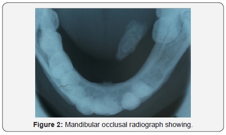
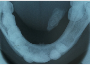
Extraoral examination revealed mild swelling in left submandibular region with no other significant findings. Upon intraoral examination a well-defined swelling with exposed hard yellowish mass of approximately 1.5x 1.0cm was found in left canine premolar region of floor of mouth. Overlying mucosa was normal in color except in the anterior most part of swelling where there was an opening with exposed yellowish mass. It was hard in consistency and slight tender on palpation (Figure 1). His left mandibular second premolar and first molar was missing.A provisional diagnosis of left Wharton’s duct sialolith was made. Mandibular occlusal radiograph was advised which showed a radiopaque mass extending back beyond the lower left first permanent molar located within the Wharton’s duct (Figure 2). A final diagnosis of left Wharton’s duct sialolith was made (Table 1).


Under local anesthesia a transorallongitudinal incision was given over the duct including the preexisting sialo oral fistula. Blunt dissection was done and sialolith was removed taking care not to injure the lingual nerve. After removal of the stone thick salivary discharge came from the incised duct which was flushed and area irrigated with saline. Primary closure of only mucosa was done. Postoperative healing was uneventful (Figure 3).

Case 2
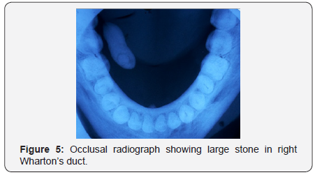
A 45 years old male reported to department of oral and maxillofacial surgery, with a chief complaint of pain and swelling while eating, in right side of floor of mouth since 1 year. There was no history of any systemic disease and patient was in good health. Extraoral examination revealed mild swelling in right submandibular region and a small fluctuant swelling in right side of chin. On intra oral examination a large, firm, nontender swelling in right floor of mouth in the region of submandibular duct was noted. Overlying mucosa was normal in color with no discharge. Mandibular occlusal radiograph was advised which showed a large radiopaque stone in right Wharton’s duct (Figures 4 & 5).

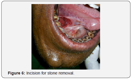
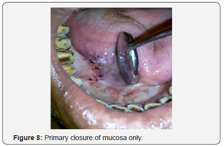
Under local anesthesia a transorallongitudinal incision was made over the duct (Figure 6). Sharp dissection was done and sialolith was removed taking care not to injure the lingual nerve. The exposed stone was grasped with the sinus forceps and gently teased but it was broken and so removed in 3 small pieces (Figure 7). After removal of the stone thick salivary discharge came from the incised duct which was flushed and area irrigated with saline. Primary closure of only mucosa was done (Figure 8). Postoperative healing was uneventful.

At a follow up period of 6 months both patients were fine.
Discussion
The submandibular gland is the site for majority of salivary calculi and this may be due to the direction of salivary flow (against gravity), long tortuous duct course, more alkaline pH and high calcium and mucin content, but the exact etiology and pathogenesis are unknown. The age in the cases reviewed ranged from 21 to 75 years with average 51.4 years. Majority of duct stone occurred in 4th to 5th decades of life which is consistent with previous literature on sialoloith. Among the 34 cases reviewed in this article the incidence is higher in male (n=28) compared to females (n=6) with male to female ratio of 4.7:1. Size of stone in the reported cases ranged from 15mm to 72mm. Stones over 10 mm are reported as unusual in size. The two cases reported here are more than 20mm in size. The largest stone reported has been presented by Rai and colleague [1] which was 72mm in size. The ability of a calculus to become giant depends mainly on the reaction of the affected duct. If the duct adjacent to the sialolith is able to dilate allowing nearly normal salivary flow, it might remain asymptomatic for a long period; thus eventually creating a giant calculus [6]. Out of 34 cases reviewed, 19 cases involved left side of duct and 11 cases involved right side duct and in 4 cases side was not mentioned. So from above data it can be predicted that sialolith of unusual size mainly occurs in left Wharton’s duct. In our cases 1 patient had sialolith in left and 1 patient had right.
In the above reviewed cases pain and swelling was the most common symptoms for which patient visited to their physician. Cause of pain and swelling of the involved salivary gland is due to obstruction of salivary flow during the food related surge [7]. The severity of symptoms depends on the degree of obstruction. In few cases patients were asymptomatic.
The diagnosis can be made by history, physical examination and using Ultrasonography, Xrays (most commonly occlusal radiographs or OPG), Sialography or CT scan. Small stones especially which are radiolucent can be diagnosed by investigations like Computed tomography, sialography or ultrasonography. Sialoendoscopy is a new, minimally invasive technique developed for direct visualization of intra‑ductal stones [8,9]. In our cases the stone was radiopaque , large size and was clearly visible on occlusal radiographs so no further investigations were needed. Large calculi especially when present in anterior part of duct may result in floor of mouth perforation and sialo oral fistula formation. In one of our cases, calculus has extruded in floor of mouth causing sialo oral fistula formation. Similar findings was reported by El Gehani, Akimoto, Patil, Huber, Shetty; whereas Paul and Chauhan reported sialo‑oral as well as sialo-cutaneous fistula caused by sialolith [10-15].
Treatment objective for salivary duct stone is to remove the stone and restore the normal salivary flow. Numerous treatment methods has been described depending upon the size & postion of stones and ranges from application of moist warm heat with gland massage, use of sialogogue, and tranoral removal to complete gland removal (sialoadenectomy) [1]. Other methods of treatment include shock wave lithotripsy, sialoendoscopy, interventional radiology, laser fragmentation and endoscopically assisted transoral removal5.Transoral removal of stone is a versatile technique and is recommended whenever stones can be palpated intraorally. This approach is without complication however care must be given no to injure lingual nerve. We successfully treated both the cases by tranoral incision. At a follow up period of 6 months both pateints were fine with normal salivary flow. A protein rich diet with lots of liquid and acid food and drinks were advised in order to prevent recurrence. The patient should be encouraged to seek early treatment for salivary calculi because long term obstruction in the absence of infection can lead to atrophy of the gland with resultant lack of secretory function and ultimately fibrosis. However, after elimination of the obstruction, the apparent resiliency of the submandibular gland results in no adverse symptoms.
Consuegra reported two cases of giant sialolithiasis within the Wharton’s duct causing unilateral absence of submandibular gland due to complete acinar atrophy.
Conclusion
Early diagnosis and treatment should be done to prevent large submandibular stone formation which if left untreated for long period may lead to complete salivary gland fibrosis and dysfunction. The ultimate surgical removal of gland due to chronic obstruction can be avoided by educating patients about the importance of hydration and oral hygiene and prompt removal following diagnosis.

Comments
Post a Comment