Root Canal Retreatment of Permanent Mandibular Canine with Two Canals - A Case Report- Juniper Publishers
Juniper Publishers- Open Access Journal of Dentistry & Oral Health
Authored by Al Dahman Yousef H
The knowledge of both the external and internal anatomy of teeth is important for adequate root canal treatment. Permanent mandibular canine usually single-rooted with single root canal. Single-rooted mandibular canine with two canals is rare. This paper describes a case of root canal retreatment of permanent mandibular canine with two root canals and one apical foramen (Type II) in single root. The clinicians must be aware of these anatomical variations and able to use variety of tools for proper diagnosis and management
Keywords: Mandibular canine, Anatomical variation, root canal retreatment
Introduction
The objectives of root canal treatment are to eliminate infection from root canal and to prevent reinfection. [1]. Failure to locate, clean, shape, or fill all root canals can lead to post-treatment disease, pain, and/or complications of treated tooth [1-3]. Therefore, a broad knowledge of the root canal anatomy and its variations is mandatory to improve the prognosis of root canal treatment [4].
Generally, mandibular canine is usually single-rooted with single root canal. [5,6] Several reports of anatomical variations of mandibular canine have been reported in the literature. The incidence of a mandibular canine with single-root and two root canals is approximately 15%. [5,7,8] Also, the incidence of mandibular canine having double-roots and two canals was reported to be up to 5% [6,9]. The present case report describes the nonsurgical root canal re treatment of a mandibular canine with single-root and two root canals.
Case Report
A 38 years old Saudi woman was reported to endodontic postgraduate clinics of Riyadh College’s Dental Hospital, Riyadh, Saudi Arabia for nonsurgical endodontic retreatment of mandibular left canine (number #33). The chief complaint was to complete the root canal retreatment that was started by an undergraduate dental student a week ago. The patient’s medical history was noncontributory.
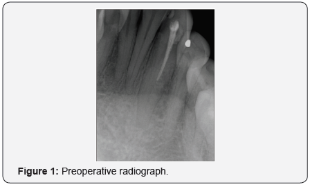
Clinical examinations revealed a temporary restoration on tooth #33. The tooth was sensitive to percussion and palpation. There was no mobility and the periodontal status was normal. Radiographic examination revealed a previously treated tooth with slight periapical radiolucency related to tooth #33 (Figure 1). Based on the clinical and radiographic findings and according to American Association of Endodontics consensus, [10] the tooth was diagnosed as previously treated with symptomatic apical periodontitis.

Following the delivery of local anesthesia (2% lidocaine and 1:100,000 epinephrine) and isolation with rubber dam, removal of the temporary filling was made. The pulpal floor was carefully examined under dental operating microscope (DOM) (Global 001 Dental Microscopes, Global Surgical Corporation, USA). Two separate buccal and lingual orifices were identified. The access cavity outline was extended buccolingually to establish straight line access.
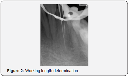
The working length was established using electronic apex locator, Root ZX II (J. Morita, Tokyo, Japan), and confirmed radiographically (Figure 2). The buccal canal joined the lingual canal in the middle third of the root (type II Vertucci’s classification) [11].

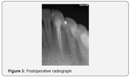
The two canals were shaped by ProTaper Next System (Dentsply Maillefer, Ballaigues, Switzerland) to size X3 for both canals. Copious irrigation with 2.5% sodium hypochlorite (NaOCl) followed by 17% ethylenediaminetetraacetic acid (EDTA) was carried out during the instrumentation phase. After the final flush, the canals were dried with paper points and obturated with matching Gutta percha cones and Endosequence BC sealer (Brasseler, Savannah, GA). The access cavity was sealed with Coltosol temporary filling material (Coltosol® F, Coltene, Switzerland), and the patient was referred to receive final restoration (Figure 3).

Discussion
Failure to locate, clean, shape, and fill a canal has been demonstrated to be a causative factor in the failure of nonsurgical endodontic therapy [12,13]. Teeth with anatomical variation are an important issue in root canal treatment. Missing root canals may contain necrotic tissue and microorganisms. Consequently, this may lead -to development of an apical periodontitis. Thus, clinicians should be aware of complex root canal structure.
Mandibular canines with single-root and single canal are common, however, two root canals, [5,7,8,14,15] and in some unusual cases, there may be one or two roots with three root canals [16,17] has been reported.
In the present case, mandibular canine with single-root and two canals was reported. The incidence of two root canals in single rooted mandibular canine teeth has been reported to be up to 6.25% [18] with a percentage of 14% of having Type II canals in mandibular canine [7].
One case report of a Saudi female patient with single-rooted lower canine with two canals was reported [15]. The incidence of two canals in mandibular canine is more in female than male as reported in the literatures [19].
Proper interpretation of conventional periapical radiographs taken in more than one angle is mandatory to detect any morphological variations of teeth [19] Additionally, using advanced diagnostic radiographic techniques such as cone beam computed tomography (CBCT) are very helpful to detect such variations if conventional radiographic techniques lack to provide obvious information and more details are required [20].
Also, using magnification tools, magnification loupes or DOM, might assist in locating and additional canals [21]. In the present case, DOM was used and helped in locating the missed canals [22- 27].
Conclusion
Although the occurrence of single-rooted mandibular canine with two canals is infrequent, the clinician should be aware of such anatomical variations in the permanent mandibular canine. Careful interpretation of radiographs along with an appropriate access cavity design and the use of magnifying tools will help in evaluation and detecting such variations. Failure to detect these variations will result in an uninstrumented canals and subsequent failure of the root canal treatment.


Comments
Post a Comment