Bone Graft Case- Juniper Publishers
JUNIPER PUBLISHERS-OPEN ACCESS JOURNAL OF DENTISTRY & ORAL HEALTH
Periodontology
Bone Graft, ridge preservation, socket preservation,
bone grafting and alveolar ridge preservation are several different
terms used in dentistry for bone grafting. The purpose of this procedure
is to preserve and prevent bone shrinkage after an extraction.
Wang and Lang reported in 2012 that “Applying the
guided bone regeneration principle using bone substitutes together with a
collagen membrane has shown clear effects on preserving alveolar ridge
height as well as ridge width”.
Back in 2012, Dr. Gordan J Christensen stated that
“socket grafting is not yet ‘standard of care,’ but in my opinion it
certainly should be”
There are various types of bone materials - autograft
which are from the patient’s own body, xenograft which is from a
different species, allograft which is from a human cadaver and alloplast
which is a synthetic material. I have only used allograft as this was
what was available in the office at the time.
Not only is socket and ridge preservation for dental
implants, it can also benefit by providing retention for dentures or
providing aesthetics for a bridge.
Most PPO insurances do not reimburse much for a
surgical extraction, let alone a simple extraction. Socket grafting can
help boost production in addition to giving the patient the benefits of
preventing bone shrinkage.
Here is my first socket and ridge preservation case:
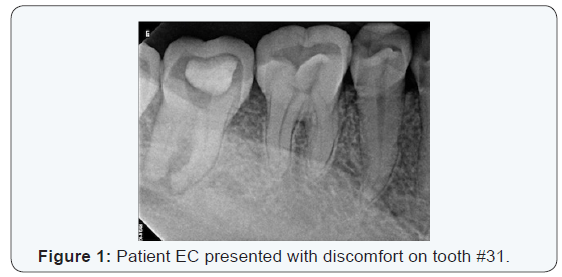
Patient EC presented with discomfort on tooth #31. Medical history
reveals he has high blood pressure and diabetes type 2 (Figure 1).

EC states his previous dentist performed a root canal
on #31. Radiograph reveals a root canal may have been initiated however
incomplete and filled with a temporary restoration. An intraoral
evaluation reveals a temporary restoration on the occlusal of #31 with
open margins. #31 also had a mobility grade of 1.
My proposed treatment plan to EC was to surgically
extraction #31 and placed a bone graft with membrane as patient
expressed interest in implants.
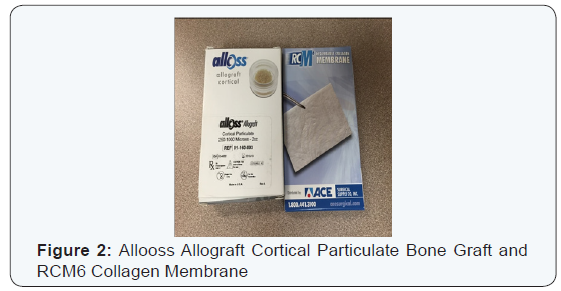
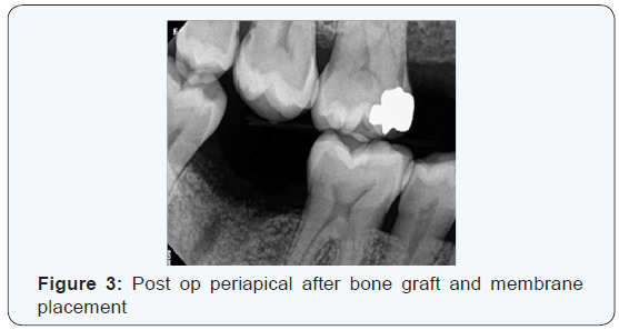
After #31 was extracted, the material available at
the time was Allooss Allograft Cortical Particulate bone graft, which
was placed into the socket and covered with RCM6 resorbable collagen
membrane. Sometimes the particulates will move out of the socket however
most of it will be covered by the soft tissue (Figure 2&3).


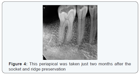
This periapical was taken just two months after the
socket and ridge preservation. Notice there is no cupping of the ridge,
which is apparent in extractions without socket and ridge preservation.
The dental director at this office commented on how great the bone graft
looked (Figure 4).

The healing time various for maxilla and mandible,
usually four to six months and two to four months, respectively. To
reduce healing time I would recommend my fellow oral physicians to
incorporate drawing the patient’s blood prior to the procedure and using
it to create a PRF (platelet rich fibrin) membrane to help shorten the
healing time.
For more Open Access Journals in Juniper Publishers please
click on: https://juniperpublishers.com
For more articles
in Open Access
Journal of Dentistry & Oral Health please click on:

Comments
Post a Comment